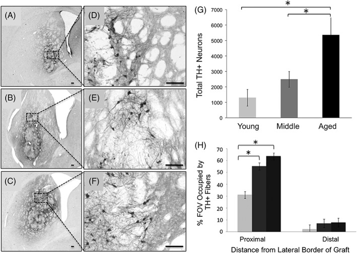
Figure 1.

The aged brain is capable of supporting survival of large numbers of grafted DA neurons and extensive graft‐derived neurite outgrowth. (A,D) DA graft in young rat; (B,E) DA graft in middle‐aged rat; and (C,F) DA graft in aged rat. (G) There are significantly more grafted TH+/DA neurons in the aged brain, (H) with significantly more TH+ fibers in the middle‐aged and aged striatum proximal to the graft compared to young striatum. Proximal: 0–850 μm from the graft border; distal: 850–1,700 μm from the graft border. Asterisks denote significant differences (P < 0.05) for indicated comparison. Images modified from Collier and colleagues.24
