Figure 3.

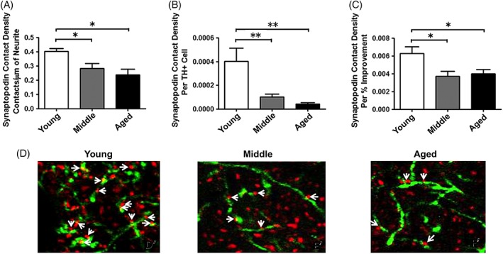
There are fewer putative synaptic contacts formed between graft‐derived TH+ neurites and synaptopodin (SP)+ dendritic spines of host MSNs in rats of more advanced age compared to young rats. SP is an actin‐binding protein located in the spine neck of MSNs. We used dual‐label fluorescence immunohistochemistry, using antibodies against TH and synaptopodin, and confocal microscopy to estimate the degree to which grafted cells in rats of varying ages were making putative synaptic connection with MSNs. Despite 5× more grafted TH+ neurons, there are significantly fewer TH/SP appositions in the aged, parkinsonian brain (A,B,D), and these appositions correlate with the degree of behavioral improvement (C). (D) TH+ neurites = green; SP+ dendritic spines = red. The asterisk (“*”) denotes P < 0.05 for the indicated comparisons. Image modified from Collier and colleagues.24 [Color figure can be viewed at wileyonlinelibrary.com]
