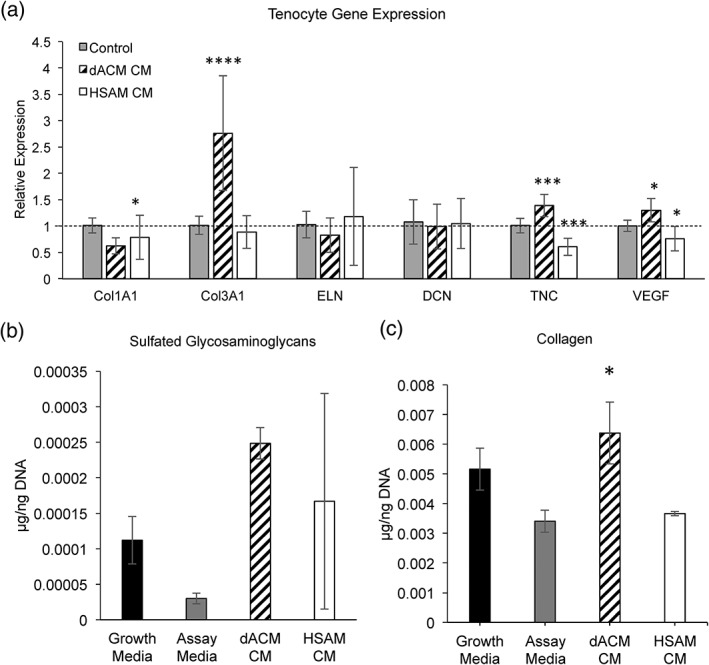
Figure 4.

Tenocyte extracellular matrix gene expression and deposition. (a) Gene expression of collagen I (COL1A1), collagen III (COL3A1), elastin (ELN), decorin (DCN), tenascin C (TNC), and vascular endothelial growth factor (VEGF). Mean ± standard deviation reported; n = 9 for all groups. * denotes p < .05; *** denotes p < .001; **** denotes p < .0001 compared with control. (b) Deposition of sulfated glycosaminoglycans (sGAGs). Mean ± standard deviation reported; n = 3 for all groups. (c) Deposition of collagen. Mean ± standard deviation reported; n = 3 for all groups. * denotes p < .05 compared with assay media. CM, conditioned media; dACM, dehydrated amnion/chorion membrane; HSAM, hypothermically stored amniotic membrane
