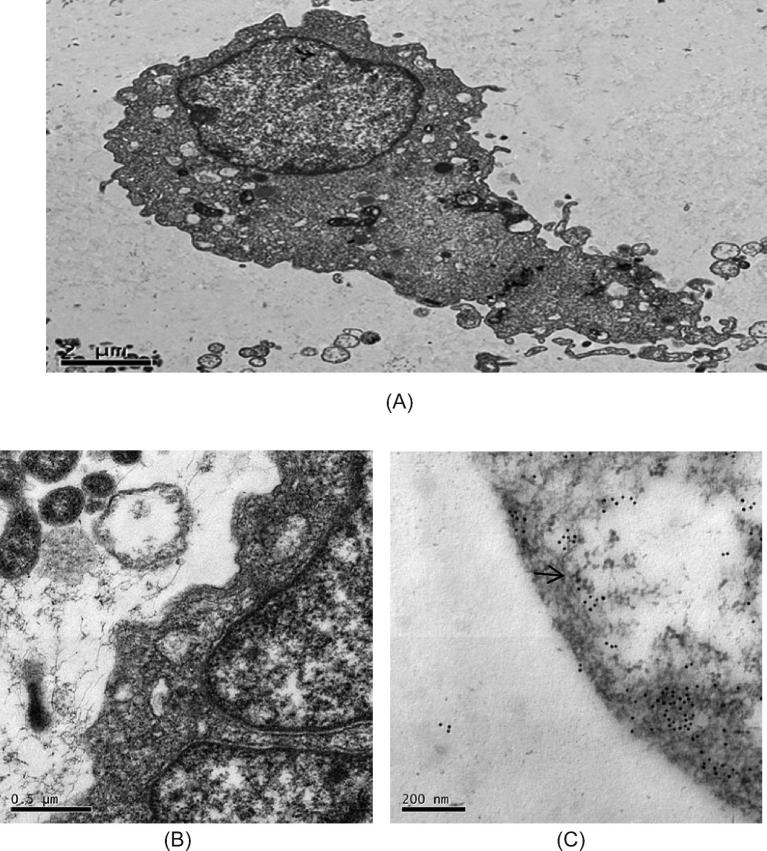
Figure 7.
Electron microscopy image of DF-1/B cells that were pre-incubated with TBS (pH7.4) and subsequently incubated with 10 nm colloidal gold-labeled goat anti-mouse IgG. These samples served as negative controls. (A) TEM × 25,000, (B) TEM × 100,000. (C) Electron microscopy image of DF-1/B cells that were pre-incubated with goat anti-mouse EGFP antibodies and monoclonal antibodies for ALV-B individually and subsequently incubated with 10 nm colloidal gold-labeled goat anti-mouse IgG. TEM × 250,000. The arrows point to gold particles.

