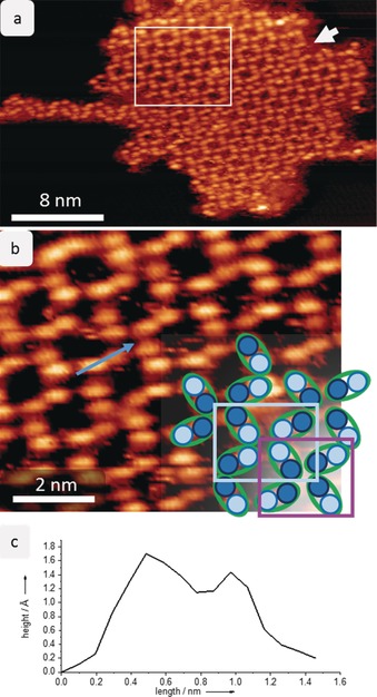
Figure 1.

Self‐assembly of sucrose on Cu(100) at 40 K. a) STM image of one island of sucrose on Cu(100) showing the periodic network. The molecules are seen as oval lobes; a white arrow indicates a domain boundary that divides the entire island. An additional structure is found on the left side of the island. The white rectangle marks the area enlarged in (b). b) Magnification of the periodic network. The sucrose molecule is imaged as double lobes with two features of different intensity. In the lower right corner, a schematic representation is overlaid to illustrate the assembly. Two motifs are formed with either bright features (light blue rectangle) or lower‐intensity features (violet rectangle) together in a node. c) The line profile on a sucrose molecule shows the height of the two different features.
