
Figure 2.
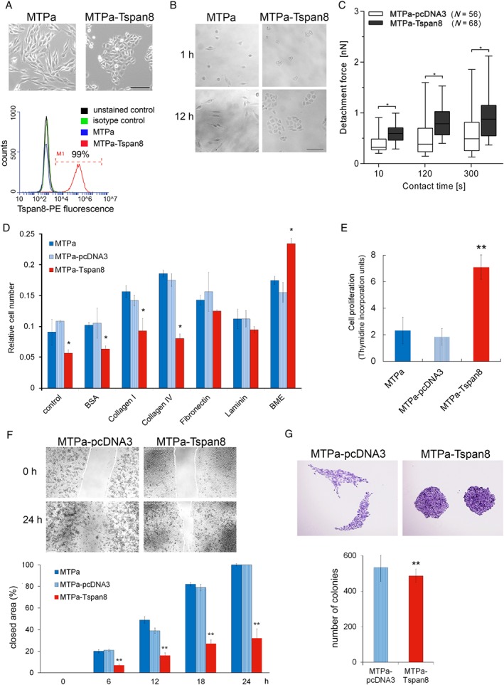
Tspan8 supports proliferation and cell–cell adhesion in breast cancer. (A) Upper panel: stable expression of Tspan8 in a rat breast cancer cell line MTPa leads to changes in cell morphology (scale bar = 200 μm). Bottom panel: Tspan8 surface expression was assessed using flow cytometry, which revealed 99% Tspan8+ cells. (B) Impact of Tspan8 on cell clustering was assessed. Cells were seeded at low density and monitored for 12 h. The experiment was repeated three times. (C) Cell–cell adhesion was tested using atomic force microscopy. Tspan8‐expressing cells exhibited significantly higher adhesion forces than cells transfected with a control pcDNA3 plasmid. The analysis on a single cell level revealed a high level of heterogeneity, showing strong variability of adhesive properties of single cells, resulting in high error bars calculated as a standard error by statistical analysis. (D) Cell–matrix adhesion was tested on different substrates using pre‐coated 24‐well plates. Tspan8 expression resulted in a significant reduction of cell–matrix adhesion when no additional coating was used and on the collagen I and IV matrixes; the BME coating resulted in a significant increase of cell–matrix adhesion of the MTPa‐Tspan8 cells. (E) To test cell proliferation, [3H]thymidine incorporation was measured. Tspan8 strongly supported cell proliferation. (F) Migration was tested using time‐lapse microscopy. Confluent cultures of the MTPa‐pcDNA3 and MTPa‐Tspan8 cells were used to produce a ‘wound’. Wound closure was observed for 24 h. Videos are available in supplementary material, Movies S1 and S2. Quantitative analysis of cell‐free areas performed using ImageJ Plugin (right panel) showed that Tspan8 strongly reduces cell migration over the ‘wound’ (scale bar = 1000 μm). The experiment was performed in biological duplicates and technical triplicates. p < 0.001 was considered as highly significant. (G) Colony‐forming ability was assessed by seeding 100 cells per 10 cm dish. While no significant difference in colony size was observed, Tspan8 mediated a slight but significant reduction of colony‐forming ability (scale bar = 500 μm).