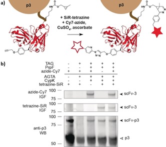
Figure 3.

Concerted dual modification of scFv–p3. a) Schematic representation of the one‐pot labeling of an scFv (PDB: 1N8Z) bearing CypK and PrpF. b) In‐gel fluorescence of scFv–p3 dually modified with tetrazine–silicon–rhodamine (tetrazine–SiR, middle, Cy5 channel) and picolylazide–sulfocyanine‐7 (azide–Cy7, top, Cy7 channel) after pulldown via the HA (human influenza hemagglutinin) tag. Western blot (bottom) probed with anti‐p3 shows p3 and scFv–p3. IGF: in‐gel fluorescence. WB: western blot.
