
Figure 1.
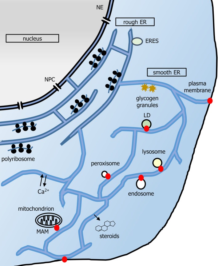
A schematic of mammalian ER. Nuclear pore complexes (NPCs) gate nucleocytoplasmic transport at the NE. The pER is composed of the rER and sER. rER (darker blue) is composed of flattened, stacked, frequently fenestrated, sheets, connected by helicoidal junctions (shown in cross section here). rER is studded with polyribosomes synthesizing secretory protein and functions in import, folding, glycosylation and onward secretion of such protein. Onward transport originates from ribosome‐free subdomains of rER (ER exit sites, ERESs). Smooth ER (sER, lighter blue) extends in a reticular network throughout the cell, characterized by three‐way junctions. ER tubules also exist in dense arrays in the perinuclear region (not shown for simplicity). The smooth ER functions in detoxification reactions, and lipid and steroid synthesis. Lipid synthesis contributes to organellar membrane generation, for example, during formation of LDs. Organelle contact sites (red dots) may also regulate signalling, for example, immune signalling and transfer of calcium to mitochondria both occur at MAMs. Contact sites may also regulate membrane dynamics, for example, endosome budding and mitochondrial fission. Although contact sites may be at the rER or sER, depending upon the organelle (e.g. both in the case of MAMs), for clarity they are depicted herein at the cell periphery. Note that the yeast ER has a different morphology; extensive cortical ER runs parallel to the plasma membrane, and is connected by tubules to the perinuclear ER, which delimits the nucleoplasm.