Figure 2.

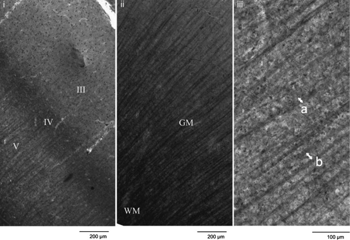
Sudan black stained section illustrating (i) cortical layers, (ii) tissue type, and (iii) measurements of axon bundle width (a) and axon bundle spacing (b), as indicated by the arrows [after McKavanagh, Buckley, & Chance, 2015]

Sudan black stained section illustrating (i) cortical layers, (ii) tissue type, and (iii) measurements of axon bundle width (a) and axon bundle spacing (b), as indicated by the arrows [after McKavanagh, Buckley, & Chance, 2015]