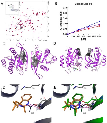
Figure 4.

NMR and X‐ray crystal structure of indolopyrrole 9 b. (A) Dose‐dependent cross peak shifts in the 2D 1H/15N HSQC NMR spectra of GCP‐KRASG12D on addition of 9 b. (B) NMR K D titration of 9 b binding to GCP‐KRASG12D. (C) Top view and (D) side view of the high‐throughput soaking crystallization system for KRASG12D with the SI/II‐pocket surface depicted in grey. (E) X‐Ray co‐crystal structure of 9 b binding to the SI/II‐pocket of GCP‐KRASG12D. (F) X‐Ray co‐crystal structure of 9 b binding to the SI/II‐pocket of GCP‐KRASG12D in yellow overlaid with the docking pose in green.
