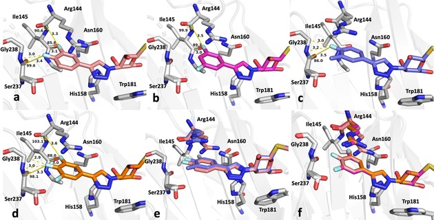
Figure 4.

Close‐up view of the fluorinated phenyl moiety in the crystal structures of galectin‐3C in complex with a) 5, b) 6, c) 7, d) 8, and e) 5 and 7. f) Superimposed structures of 5, 6, and 8 show nearly identical modes of binding. Key fluorine interactions described in the text are indicated with yellow dashed lines. Distances and F⋅⋅⋅C=O angles a2 are shown in panels a)–d).
