
Fig. 1.
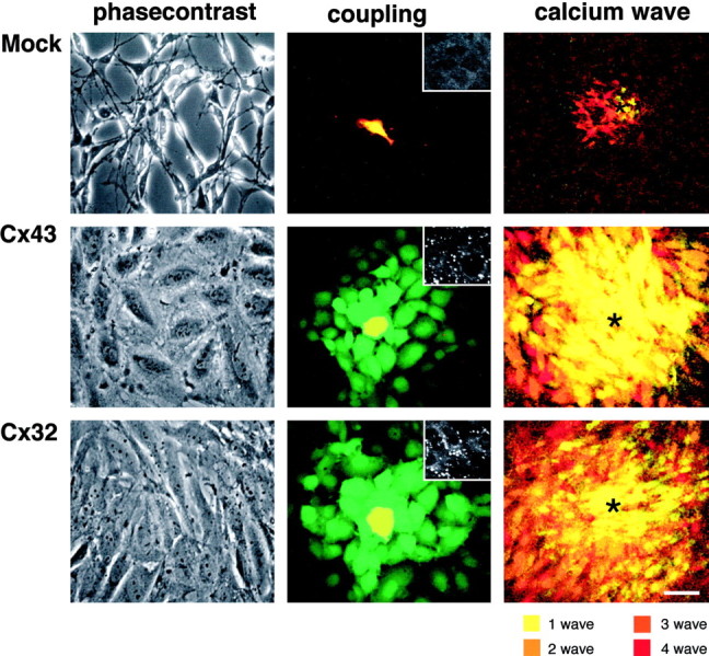
Cx expression is associated with an increase in functional coupling and a transformation of cellular phenotype. Morphology and functional coupling of mock-, Cx43-, and Cx32-transfected C6 cells are shown. Left panels, Phase-contrast micrographs of representative cultures. Control or null-transfected cultures are composed of elongated compact cells with little cell–cell contact. In contrast, both Cx43 and Cx32 cells are large, flat cells with an abundance of cellular contacts. Middle panels, Gap junctional coupling demonstrated by transfer of CDCF (green) from DiIC18-labeled cells (red) to unlabeled identical cells. Donor cells appear yellow because of the merge of red and green labeling. The mock-transfected clone is completely devoid of gap junctional coupling, whereas both C6-Cx43 and C6-Cx32 cells are extensively coupled. Insets, Cx43 immunoreactivity in mock and C6-Cx43 clones and Cx32 immunoreactivity in C6-Cx32 cells.Right panels, C6 cells acquire the ability to propagate long-distance calcium waves after Cx transfection. Four representative waves in control mock-transfected and C6-Cx43 or C6-Cx32 cells were color coded and digitally superimposed to demonstrate the extent of calcium signaling after mechanical stimulation in each transfectants (asterisks indicate point of stimulation). Scale bar:left and middle panels, 30 μm;right panel, 60 μm; insets, 15 μm.