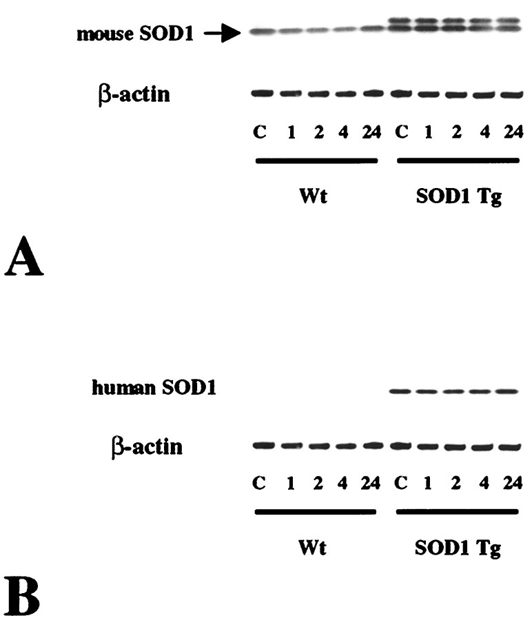
Fig. 1.
Western blot analysis of mouse (A) and human (B) SOD1 expression in wild-type (Wt) and SOD1 Tg mice before and after transient FCI. Protein (6.5 μg) from the cytosolic fraction was loaded per lane. Constitutive expression of endogenous mouse SOD1 was seen in both the wild-type and Tg mice and was not modified after FCI (A, top panel). Human SOD1 was also detected by anti-mouse antibody in SOD1 Tg mice (A, top panel), as shown by thetop bands, but not in the wild-type mice. Human SOD1 was not detected in the wild-type mice, but it was detected in the SOD1 Tg mice before and after FCI. There was no change in the human SOD1 level before and after FCI in the SOD1 Tg mice (B, top lane). A consistent amount of β-actin expression is shown in the bottom lanes. C, Control.

