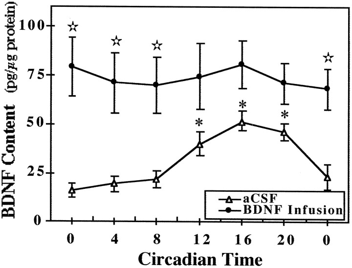
Fig. 3.
Temporal pattern of BDNF content in the SCN of rats receiving infusion of aCSF or BDNF. Symbolsrepresent the mean values (±SEM) of BDNF protein expression in the SCN of rats that were infused with aCSF or BDNF (250 ng/d) for 14 d and then killed at 4 hr intervals (n = 4) in DD. At CT 12, 16, and 20, BDNF levels in aCSF-infused rats were significantly greater (*p < 0.05) than those observed at all other times. Significant differences in the SCN content of BDNF (⋆p < 0.05) were observed between aCSF- and BDNF-infused rats at CT 0, 4, and 8.

