Fig. 1.
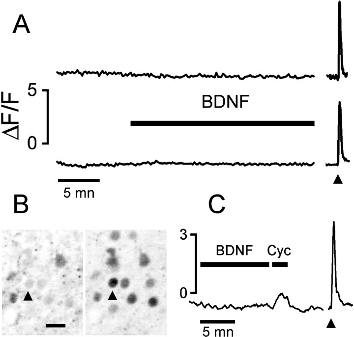
BDNF did not cause any detectable change of [Ca2+]i in neurons of cortical slices.A, Fluorescence from the Ca-indicator fluo-3 was integrated at the cell body of two neurons from a P8 and a P18 rat (top and bottom traces, respectively). The arrowhead indicates the stimulation with 20 μm NMDA for 10 sec. B, The field is shown as recorded during the BDNF (200 ng/ml) presentation (left) and at the peak of the NMDA response (right). The arrowhead points to the cell for which fluorescence is plotted on the top trace. Images are presented as negatives for better clarity. Scale bar, 20 μm. C, The lack of response to BDNF is not attributable to low sensitivity of the imaging because the small calcium increase induced by release from intracellular stores was easily detectable. Cyclopiazonic acid (Cyc; 50 μm) caused a small but clearly resolved calcium transient in 49% of neurons (n = 57), whereas BDNF presentation did not cause any detectable calcium change.Arrowhead points to a 10 sec puff of 20 μmNMDA.

