Fig. 5.
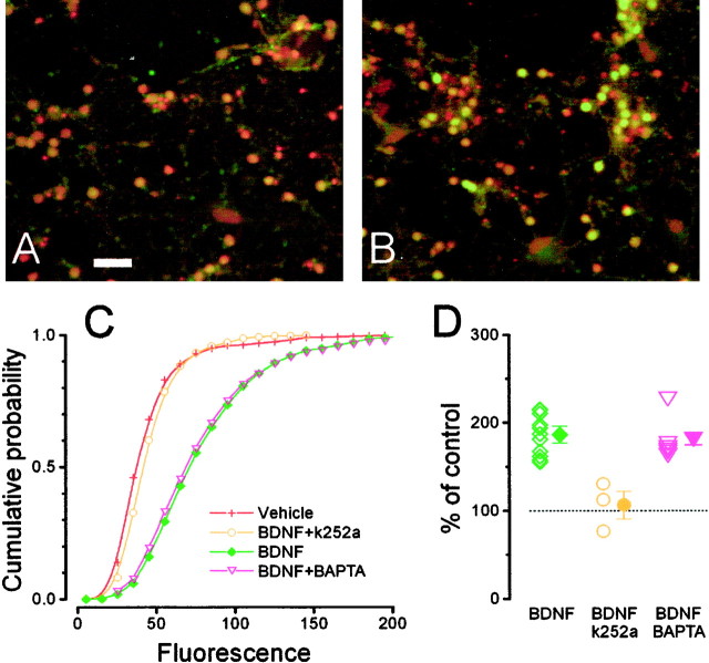
Stimulation with BDNF (1 hr) caused calcium-independent c-fos expression in cultured neurons from the visual cortex through a Trk-dependent pathway. A, Double staining with the nuclear staining TOTO (red) and a c-fos antibody (green). In control conditions only a weak green fluorescence is present in the neuron nuclei. Scale bar, 50 μm. B, After incubation with 50 ng/ml BDNF, most neurons became intensely positive for c-fos. C, The cumulative probability of the fluorescence distribution shows that BDNF (3543 cells) caused c-fos expression (green) and that this effect is completely suppressed by incubation with the Trk inhibitor k252a (k252a, 964 cells, yellow; vehicle, 2374 cells, red; one-way ANOVA, p < 0.005; Tukey's post hoc test, BDNF vs vehicle,p < 0.05; k252a vs vehicle, p> 0.05). Loading neurons with BAPTA does not affect BDNF-induced c-fos expression (BDNF + BAPTA, 1523 cells, magenta; Tukey'spost hoc test, p > 0.05).D, Mean ± SEM and single results for all experiments. BAPTA alone did not modulate c-fos expression (92.8 ± 2.5% of control, 1021 cells; data not shown in figure).

