
Fig. 5.
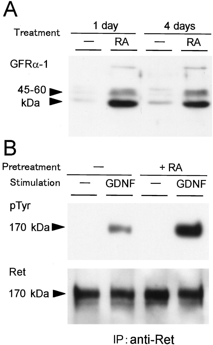
Effects of the RA treatment on the levels of GFRα-1protein and GDNF-induced Ret tyrosine phosphorylation in cultured SCG neurons. A, SCG neurons were treated without or with 10−7m RA in the presence of 40 ng/ml NGF for 1 or 4 d before cell lysate was prepared. The cell lysate (7 μg of protein) was subjected to Western blotting with anti-GFRα-1 antibody. B, SCG neurons were pretreated without or with 10−7mRA in the presence of 40 ng/ml NGF for 4 d. Then, SCG neurons were stimulated with 50 ng/ml GDNF for 5 min before cell lysate was prepared. The cell lysate was immunoprecipitated with anti-Ret antibody and subjected to Western blotting with anti-phosphotyrosine antibody (top panel). Blotted membrane was reprobed with anti-Ret antibody to evaluate the amounts of recovered Ret protein (bottom panel).