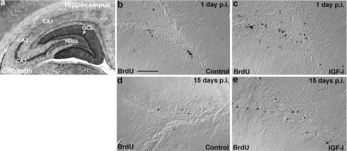
Fig. 1.
BrdU immunohistochemistry in two experiments in which IGF-I was given for 6 d (corresponding to 1 d p.i. of BrdU) or 20 d (corresponding to 15 d p.i. of BrdU). a, The hippocampal region of the adult rat brain immunoperoxidase-stained for the neuronal marker Calbindin D28K. b–e, Differential interference contrast photomicrographs of BrdU-immunopositive cells in the hippocampus. A comparison of BrdU labeling in controls (b, d) and IGF-I-treated animals (c,e) at 1 and 15 d p.i. of BrdU is shown. (For quantification, see Fig. 2.) Scale bar, 100 μm.

