
Fig. 2.
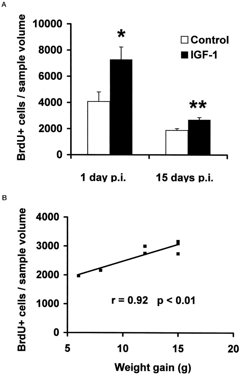
Quantification of BrdU-positive cells in the adult rat hippocampus. A, The density of BrdU-positive cells (cells per cubic millimeter of sample volume) in the GCL 1 and 15 d p.i. was determined stereologically. IGF-I-treated animals (n = 4 at 1 d p.i.; n = 7 at 15 d p.i.) are hx rats given l-thyroxine, cortisol, and IGF-I, as described in Materials and Methods. Controls (n = 5 at 1 d p.i.; n = 8 at 15 d p.i.) are hx rats given l-thyroxine and cortisol only. Means ± SEM are given. *p < 0.05; **p < 0.01. B, Weight gain and the number of BrdU-labeled cells in the GCL at the individual level are compared. There is a correlation between S-IGF-I and BrdU-positive cells after 20 d of IGF-I treatment (15 d p.i.); animals with a relatively low serum IGF-I have relatively low numbers of BrdU-positive cells, and vice versa.