
Fig. 3.
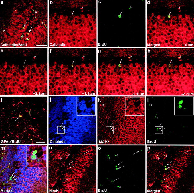
Colocalization of BrdU immunoreactivity (green–yellow in a, c–i, l, m, o, p) with immunoreactivity of the granule cell marker Calbindin D28K (red in a–h;blue in j,m), the astrocyte marker GFAP (red in i), the neuronal marker MAP2 (red in k,m), or the neuronal marker NeuN (red inn,p) (arrows indicate colocalization in a–p). Red blood cells and endothelial cells in several small blood vessels also emit nonspecificgreen and red fluorescence (asterisks in a–p). The specificity of BrdU and Calbindin D28K coexpression in three dimensions is demonstrated by a Z-series of focal planes above (e,f) and below (g,h) the focal plane shown in d(arrows in e–h indicate the same cell as in d). The merged image of b andc is shown in d, the merged image ofj, k, and l is shown inm, and the merged image of n and ois shown in p. Insets of eachboxed area in j–m are magnified 2.5×. Scale bars: a, i, 50 μm; b—h,j—m, n—p, 25 μm.