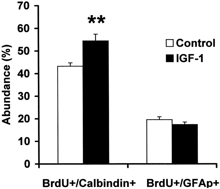
Fig. 4.
Percentage of the surviving BrdU-positive cells after 20 d of IGF-I treatment that differentiated into either neurons or astrocytes. Colocalization of BrdU immunoreactivity with cell-specific markers, either the granule cell marker Calbindin D28K or the astrocyte marker GFAP, was monitored to determine the phenotype of newborn cells after treatment with IGF-I, when compared with controls in the GCL (n = 5 for controls; n = 7 for IGF-I-treated animals). Means ± SEM are given. **p < 0.01.

