Fig. 4.
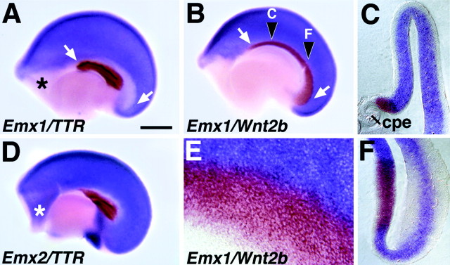
Nested expression of Emx1 andEmx2 at E12.5 defines the cortical hem.A, B, D, E12.5 cerebral hemispheres from wild-type mice, processed as whole mounts for two-color in situ hybridization, viewed from the medial side. Rostral is to the left. E, A higher magnification view of one such hemisphere. C,F, Coronal sections through a hemisphere at the rostrocaudal levels marked in B. A,D, The choroid plexus epithelium (cpe) is marked by expression of TTR (brown). Emx1expression (blue in A) avoids a band of cortical neuroepithelium curving dorsal and caudal to the cpe (arrows) and a rostral telencephalic region (asterisk). Emx2 (blue inD) is expressed in both of theseEmx1-poor regions. Emx2 is not expressed in the cpe or in a narrow strip of junctional epithelium (white in D) adjacent to the cpe (D). B, C,E, F, Wnt2b expression (brown) fills the curving band of cortical neuroepithelium that is Emx1-poor (B,C, E, F) and defines the cortical hem. At the boundary of the cortical hem,Wnt2b and Emx1 expression may overlap by one or two cell widths, best seen at higher magnification (E). The rostral telencephalic region that is also Emx1-negative (asterisks inA and D) is not part of the cortical hem. Scale bar: A, B, D, 500 μm; E, 65 μm; C, F, 170 μm.

