
Fig. 1.
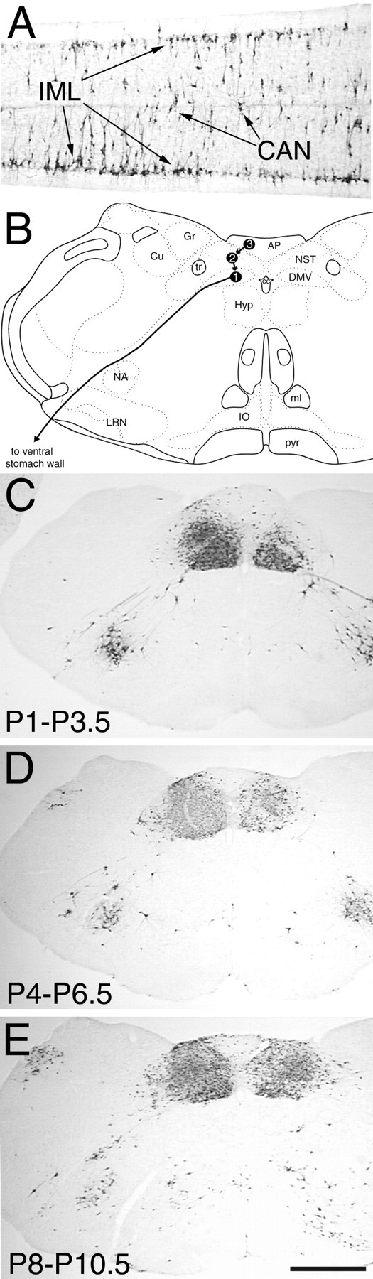
PRV immunolabeling in the thoracolumbar spinal cord (A) and caudal brainstem (C–E) 62–64 hr after inoculation of the ventral stomach wall in rats on P1 (A, C),P4 (D), or P8(E). B, A schematic diagram of the caudal brainstem [modified from Swanson (1992)] indicating the temporal progression of PRV retrograde transneuronal infection from the stomach wall to preganglionic motor neurons in the DMV(1), to preautonomic neurons in theNST (2), and finally to neurons in the AP (3), as determined in previous work (Rinaman et al., 1999). AP, Area postrema;CAN, central autonomic nucleus; Cu, cuneate nucleus; DMV, dorsal motor nucleus of the vagus;Gr, gracile nucleus; Hyp, hypoglossal motor nucleus; IML, intermediolateral cell column;IO, inferior olive; LRN, lateral reticular nucleus; ml, medial lemniscus;NA, nucleus ambiguus; NST, nucleus of the solitary tract; P1, postnatal day 1;pyr, pyramidal tract; tr, solitary tract. Scale bar: C–E, 1 mm; A, 200 μm.