
Fig. 3.
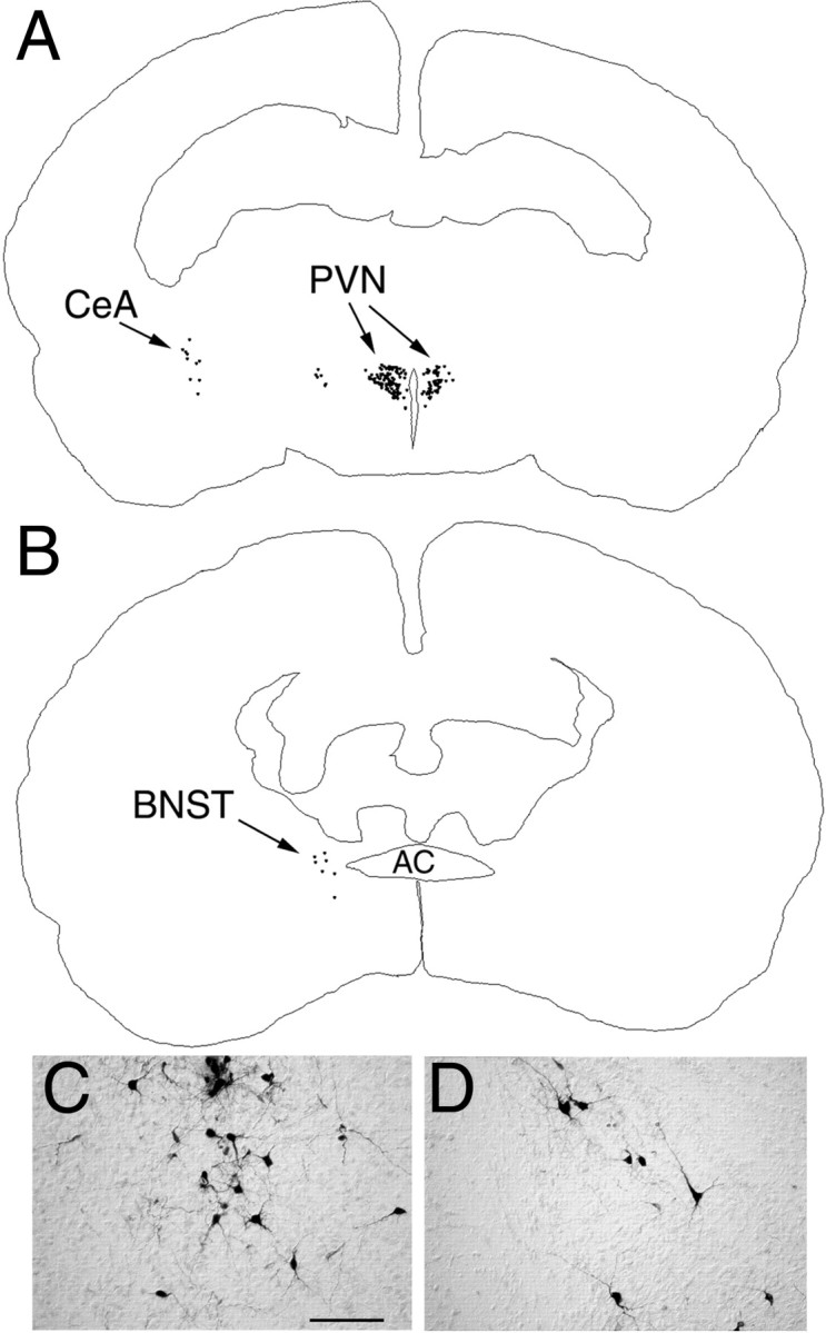
PRV immunolabeling in the diencephalon and telencephalon 62–64 hr after inoculation of the ventral stomach wall in rats on P4. A, B, Computer-assisted tracings of single tissue sections in which the distribution of all PRV-labeled neurons is plotted. C, Photomicrograph of PRV-positive neurons in the medial CeA. D, Labeled neurons in the dorsolateral BNST. AC, Anterior commissure; BNST, bed nucleus of the stria terminalis; CeA, central nucleus of the amygdala;PVN, paraventricular nucleus of the hypothalamus. Scale bar, 200 μm.