
Fig. 5.
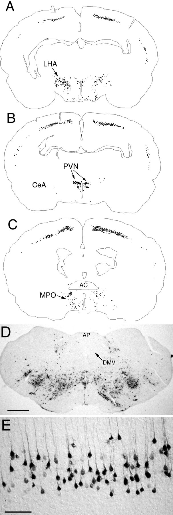
PRV immunolabeling in the diencephalon and telencephalon 62–64 hr after inoculation of the abdominal muscles in control rats on P8. A–C, Computer-assisted tracings of single tissue sections in which the distribution of all PRV-labeled neurons is plotted. D, Photomicrograph of PRV-positive neurons in the caudal brainstem. E, Labeled neurons in the primary motor cortex (layer V). AC, Anterior commissure; AP, area postrema;CeA, central nucleus of the amygdala;DMV, dorsal motor nucleus of the vagus;LHA, lateral hypothalamic area; MPO, medial preoptic area; PVN, paraventricular nucleus of the hypothalamus. Scale bars: D, 500 μm;E, 200 μm.