
Fig. 6.
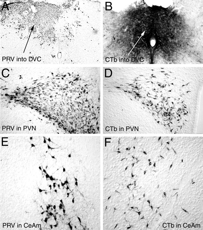
Photomicrographs of PRV andCTb immunolabeling 48–50 hr after injection of either tracer directly into the DVC on P1. A, B, Typical PRV (A) orCTb (B) injection sites in the caudal dorsomedial medulla. C, D, RetrogradePRV (C) or CTb(D) labeling in the PVN. E, F, Retrograde PRV (E) orCTb (F) labeling in the medialCeA. CeA, Central nucleus of the amygdala, medial (m); CTb, cholera toxin β subunit; DVC, dorsal vagal complex;PRV, pseudorabies virus; PVN, paraventricular nucleus of the hypothalamus.