
Fig. 3.
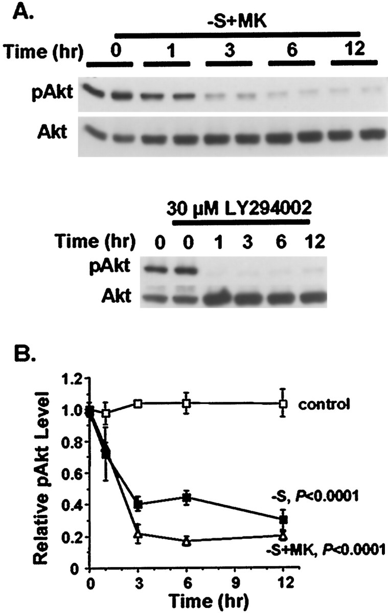
Inhibition of the PI-3 kinase–Akt pathway after trophic support withdrawal. Cortical neurons were treated for the indicated times with 30 μm LY294002, serum deprivation (−S), or serum deprivation with exposure to 10 μm MK-801(−S+MK). Neurons washed similarly but then placed in serum-containing conditioned media were used as controls (control). A, Representative phospho-Akt Western analysis (pAkt) demonstrating reduced Akt phosphorylation at Ser-473, indicative of PI-3 kinase inhibition. The blots were reprobed to show that the total level of Akt (Akt) remained constant. B, Quantitation of Akt phosphorylation by densitometric analysis of pAkt Western blots. Serum withdrawal and serum withdrawal together with MK-801 significantly decreased Akt phosphorylation compared with control washed neurons (p < 0.0001, ANOVA). Data represent averages of duplicate determinations in three independent experiments. Error bars are SEM.