
Fig. 2.
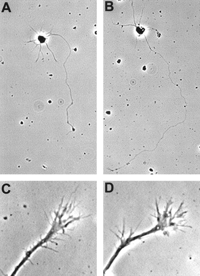
Morphology of rat cortical neurons expressing XAC(wt). Typical isolated neurons are shown, which were grown in culture for 3 d and were either not infected (A) or infected with the XAC(wt)-expressing recombinant adenovirus at 150 pfu/cell (B). Neurons shown were from fixed, low-density cultures. Growth cones of neurons infected with the recombinant adenoviruses had motile growth cones with dynamic lamellipodia and numerous labile filopodia (C, D). Growth cones are shown from presumptive axons of pyramidal neurons 3 d after infection with the GFP-expressing (C) or the XAC(wt)-expressing (D) adenoviruses. Images were taken of growth cones from live neurons.