
Fig. 5.
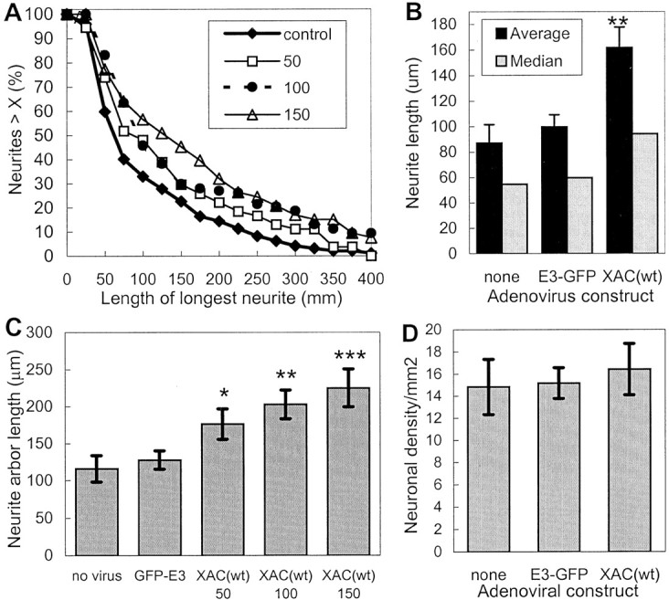
Increased neurite extension induced by the expression of XAC(wt). The length of the longest neurite was measured for rat cortical neurons. All analyses were performed blind of the experimental treatment and taken from a single experiment.A, The length distribution is shown from neurons infected with adGFP-XAC(E3) at 100 pfu/cell (control) or increasing multiplicities of infection with adXAC(wt) (shown in the legend as plaque-forming units per cell). Infection at 50 pfu/cell increased neurite lengths, as shown by a shift to the right of the length distribution curve. Infection at 150 pfu/cell increased lengths even further, with the length distribution curve from neurons infected at 100 pfu/cell falling in between or overlapping that of cultures infected at 50 or 150 pfu/cell. B, Average and median neurite lengths are shown for uninfected cultures and cultures infected with GFP-XAC(E3) and XAC(wt)-expressing adenoviruses at 100 pfu/cell. C, The total arbor length, measured by tracing the length of all branches on the longest neurite, is shown for cultures infected with control adenoviruses (100 pfu/cell) or varying MOIs of adXAC(wt). Statistically significant differences between GFP-E3 and XAC-infected cultures are indicated (*p < 0.05; **p < 0.01; ***p < 0.01; t tests).D, Infection with the adenoviruses did not affect cell densities. Measures were taken from eight randomly selected 1.2 mm2 areas/treatment. Error bars indicate SEM.