
Fig. 8.
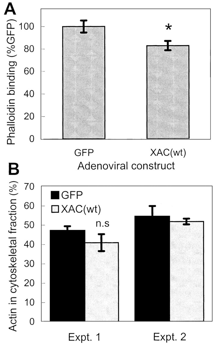
Changes in F-actin levels in neurons expressing XAC(wt). A, Three days after recombinant adenovirus infection, rat cortical cultures were fixed and F-actin was stained with Texas Red-phalloidin. Peak fluorescence intensity measured in growth cones indicated a decrease in phalloidin binding in adXAC(wt)-infected neurons compared with adGFP-infected controls (n > 30; *p < 0.05).B, A second method to determine F-actin levels was to use Triton X-100 to extract soluble actin and compare this with levels of actin in the Triton-insoluble, F-actin-containing fraction. Results from two separate experiments are shown (Expt. 1,n = 4 per group; Expt. 2,n = 2 per group). Differences were not statistically significant (n.s.) in experiment 1.