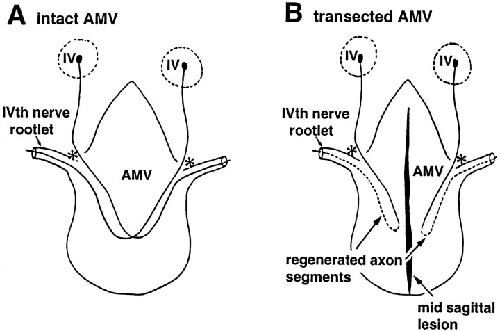
Fig. 1.
Schematic representation of whole mounts of rat AMV. A, Intact; B, transected by a midsagittal penetrant lesion. In the intact specimen, trochlear fibers from the ipsilateral IVth nucleus (IV) in the midbrain invade the AMV at the entry point (*); the majority decussate and exit in the contralateral IVth nerve rootlet (A). Midsagittal transection of the crossing axons precipitates degeneration of all severed distal segments coursing contralaterally from the midline into each rootlet (B). From the severed proximal stumps of IVth nerve fibers, regenerating sprouts grow into the ipsilateral halves of each velum, specifically directed into each ipsilateral IVth nerve rootlet, represented as dashed lines. Modified fromBerry et al. (1998).

