
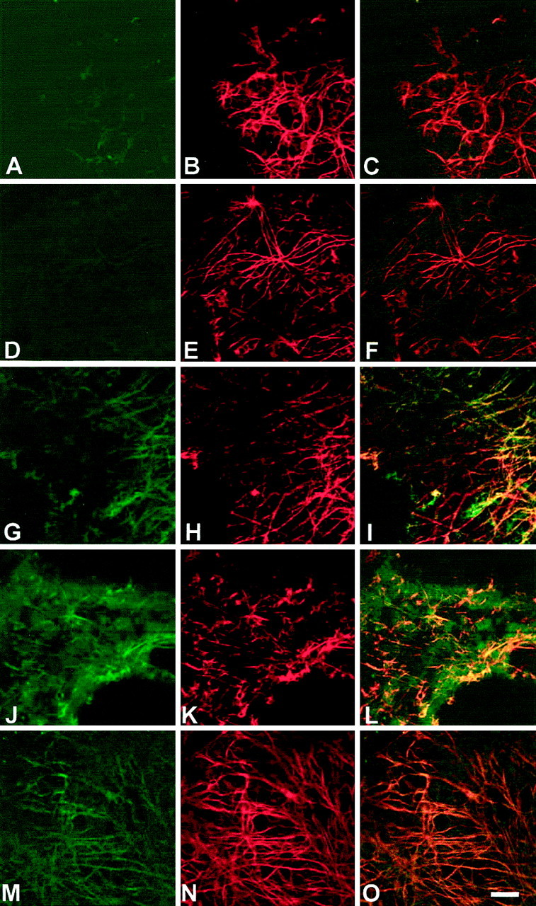
Fig. 2.
Thrombomodulin protein increases on astrocytes after lesion of the AMV. Double-immunolabeling by thrombomodulin (left) and GFAP (middle) of astrocytes of whole mounts of AMV at different times after lesioning as observed by confocal microscopy. Times after lesion are as follows: 0 hpl (A–C); 9 hpl (D–F); 1 dpl (G–I); 2 dpl (J–L); and 6 dpl (M–O). Immunohistochemistry was performed with monoclonal 3E2 anti-thrombomodulin and polyclonal anti-GFAP antibodies, which were revealed by fluorescein isothiocyanate-conjugated (green) and tetra-methylrhodamine-conjugated (red) secondary antibodies, respectively. Colocalization of thrombomodulin and GFAP on astrocytes is shown by yellow (right), maximal at 2 dpl. Pictures represent a representative sample of five experiments. Scale bar, 50 μm.