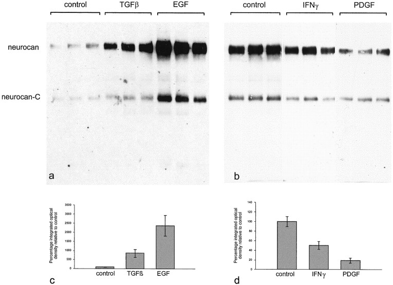
Fig. 11.
Quantification of the effects of TGFβ, EGF, IFNγ, and PDGF on neurocan expression in astrocytes. Three flasks of astrocytes were grown in the presence of each cytokine (10 ng/ml) for 3 d. The conditioned medium was treated with chondroitinase ABC, and an equal amount of protein (50 μg in a; 200 μg in b) was applied to each lane. The blot was labeled with the anti-neurocan mAb 1G2. The amount of neurocan core protein in each lane was quantified by densitometry. TGFβ brought about a ninefold increase in the amount of neurocan detected, and EGF caused a 23-fold increase (c). PDGF reduced neurocan levels to ∼20% of control, and IFNγ brought about a 50% reduction (d). The error bars represent SEM. By Student'st test, the effects of TGFβ, EGF, and PDGF were significant with p < 0.01. The effects of IFNγ were less significant (p < 0.05).

