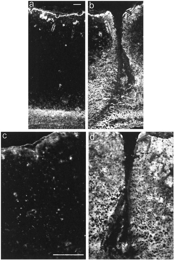
Fig. 2.

Immunolocalization of neurocan in a CNS lesion. Coronal frozen sections were labeled with the anti-neurocan mAb 1G2 7 d after a knife lesion to the cerebral cortex. The images ina and b were taken with a 10× objective, and those in c and d were taken with a 20× objective. The dorsal surface of the brain isuppermost. Labeling is apparent around the lesion (b, d), which is clearly lacking on the uninjured side (a, c). Scale bar, 100 μm.
