
Fig. 2.
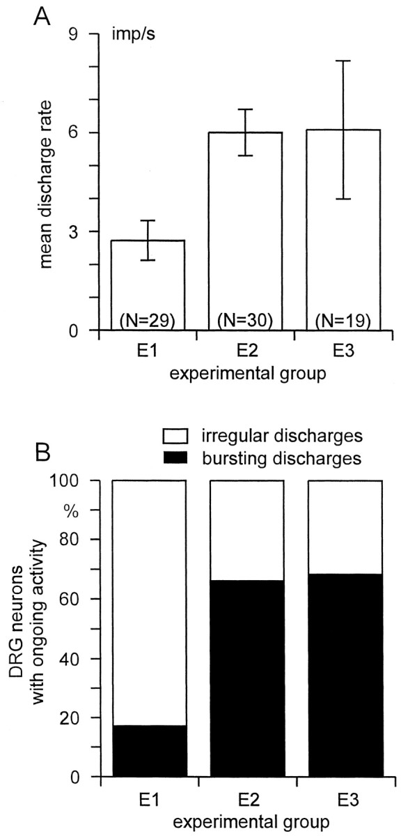
A, Mean discharge rate (±SEM) of spontaneously active DRG neurons with myelinated axons projecting into the GS nerve. B, Prevalence of different types of discharge pattern among DRG neurons that was significantly different between E1 and E2/E3 (p < 0.001, χ2 test). Experimental groups: E1, GS and sural nerve cut;E2, tibial, peroneal, sural, and GS nerve cut;E3, GS nerve intact, tibial, peroneal, and sural nerve cut.