Fig. 4.
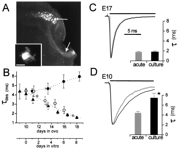
Culture environment arrests the developmental switch to fast AMPA receptor kinetics. A, Fluorescent image of contralateral (uninjected) side of slice after retrograde labeling of nMag neurons with TMR. Arrow, Labeled fibers in dorsal acoustic commissure. Double arrow, Labeled nMAG cell bodies in situ. Inset, Representative fluorescent E11 neuron retrogradely labeled and acutely dissociated. Scale bar, 10 μm. B, AMPA receptor desensitization time constant from neurons in slices (filled triangles; data from Fig. 1), acutely dissociated TMR-labeled neurons (open circles;n = 3–9 cells), unlabeled acutely dissociated neurons (gray filled circles;n = 8–9), and neurons cultured from E10 embryos (black filled circles; n = 9–16).C, Representative scaled traces of desensitizing currents in patches from E17 neurons either acutely dissociated (gray trace) or grown for 14–16 d in culture (black trace). Ten millimolar glutamate was applied during the time period marked by the horizontal bar.Inset shows average desensitization time constants of acutely dissociated (n = 10) or cultured E17 (n = 22) neurons. Time constants were not significantly different (p = 0.49).D, Scaled traces from E10 neurons either acutely dissociated (gray trace) or grown for 14–16 d in culture (black trace). Inset shows average desensitization time constants of acutely dissociated (n = 6) or cultured E10 (n = 20) neurons. *p < 0.05 indicates significance.

