
Fig. 2.
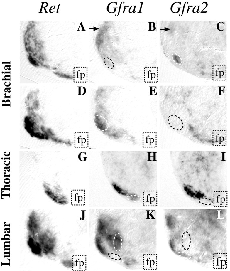
Gfra1 is more widely expressed in motoneurons than Gfra2. Whole-mount ISH was performed on E13.5 spinal cords that were subsequently sectioned. Transverse sections were performed at brachial (A-F), thoracic (G–I), or lumbar (J–L) levels. In the brachial region sections were performed either at the level of the yellow circle (A–C) or thewhite circle (D–F) drawn on Figure 1. Panels show half-ventral horn of each section;fp indicates the floor plate. Nearly all motoneurons are stained using a Ret probe (A, D, G, andJ), whereas a Gfra1 probe (B, E, H, and K) stained more motoneurons than hybridization using a Gfra2 probe (C, F, I, and L). Arrowsin B and C indicate the sameGfra1- and Gfra2-positive column as in Figure 1, C and D. Dashed lines delineate motoneuron groups that are positive for only one α-receptor.