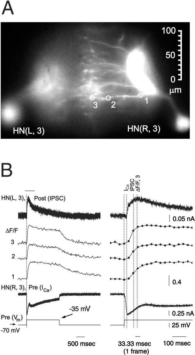
Fig. 10.

Presynaptic low-threshold Ca currents, presynaptic changes in Ca fluorescence, and associated IPSCs are correlated in heart interneurons. A, Image of preparation (dorsal side up) showing sites for recording changes in normalized Ca fluorescence (ΔF/F). B, Simultaneous recordings of presynaptic low-threshold Ca currents (ICa) and changes in Ca fluorescence and IPSCs in voltage clamp from a pair of oscillator interneurons. Traces on the right are expanded sections of the traces on the left marked by the bar. The preparation was bathed in 0 mm Na+/5 mmCa2+ saline and repeated (n = 4; average traces shown) depolarizing voltage pulses to −35 mV (from a holding potential of −70 mV) were imposed on the presynaptic cell. The postsynaptic cell was held at −40 mV. Changes in Ca fluorescence (ΔF/F) recorded at 30 Hz. Dotted lines show the start of presynaptic depolarizing step and the 90% maximal value of presynaptic ICa, Ca fluorescence in zone 3, and IPSC. Same preparation as in Figure 4.
