
Fig. 2.
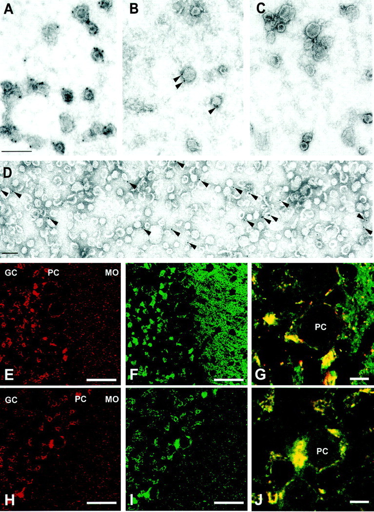
VGAT is present on a subset of synaptic vesicles. Immunogold labeling of synaptic vesicles purified through controlled-pore glass chromatography using anti-synaptophysin antibody (poly-clonal) (A), anti-VGAT antibody (B), and preimmune serum (C). D shows an overview of VGAT labeling at a lower magnification. Counting of labeled vesicles from several independent experiments revealed that ∼16% of all small vesicular profiles were labeled with anti-VGAT antibody, whereas synaptophysin labeling was observed on virtually all synaptic vesicles. Scale bars: A, D, 100 nm. Comparison of the staining patterns for VGAT (E,H) with those of synapto-brevin 2 (F) and GAD (I) in sections of rat cerebellum. The staining patterns of VGAT and GAD are identical. In contrast, synaptobrevin 2 antibody stains many more nerve terminals than VGAT; this is particularly obvious in the molecular cell layer (MO). High-magnification overlays of VGAT (red) and GAD (green) (J), and VGAT (red) and synaptobrevin 2 (green) (G) showing Purkinje cell bodies. GC, Granular cell layer;PC, Purkinje cell layer; MO, molecular cell layer. Scale bars: E, F,H, I, 50 μm; G,J, 10 μm.