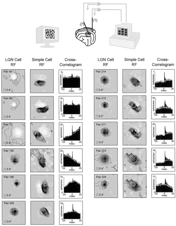
Fig. 1.

Simultaneous recordings from geniculate cells and simple cells with overlapping receptive fields. Top, Illustration of the experiment. A single electrode was inserted into layer 4 of visual cortex; up to seven electrodes were inserted into the LGN. Visual responses to a white-noise stimulus (shown) and drifting sine-wave grating were recorded. Bottom, Receptive fields and cross-correlograms for each pair of cells (n = 11) that were monosynaptically connected and studied for interactions between geniculate spikes. Regions of a cell's receptive field excited by the bright phase of the white-noise stimulus (see Materials and Methods) are shown in white; regions excited by the dark phase are shown in black. The two panels for each pair of receptive fields correspond to the same stimulus window.Circles represent a Gaussian fit to the geniculate receptive field (radius: 2.5ς). Stimulus pixel size was 0.4°. The cross-correlograms shown to the right of the receptive fields each have a short-latency peak (above the stimulus-dependent shuffle correlogram, shown in gray), indicating a monosynaptic connection between the geniculate cell and the simple cell.
