
Fig. 11.
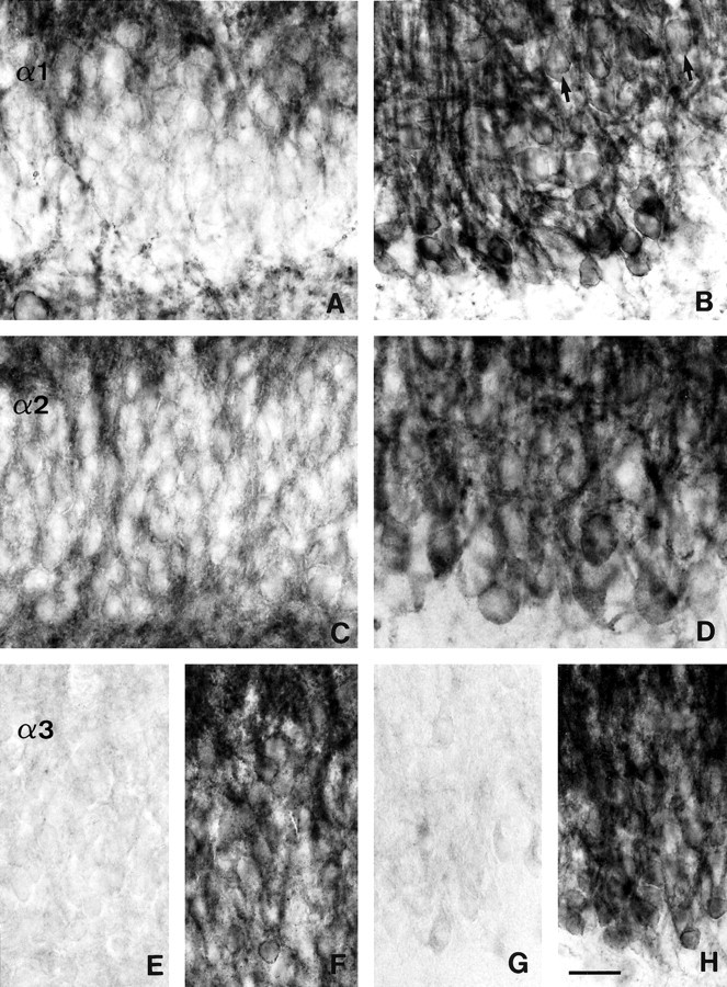
Changes in subunit-specific GABAAreceptor immunoreactivity in the dentate gyrus in TLE with HS (B, D, G, H) versus controls (A, C, surgery, TLE without HS; E, F, autopsy).A, B, α1-subunit; C, D, α2-subunit;E–H, α3-subunit. A, C, In control specimens, the somata of granule cells are outlined by faint or no staining for the α1-subunit (A) and the α2-subunit (C), whereas apical and basal dendritic fields are moderately labeled. B, D, In TLE with HS specimens, strongly increased α1-subunit (B) and α2-subunit immunoreactivity (D) is seen surrounding the somata of granule cells and in the molecular layer, whereas only few basal dendrites remain. Note the granule cell dispersion into the molecular layer in HS (B, arrows). E, F, Two control specimens illustrate the range of α3-subunit immunoreactivity. Whereas inE staining is absent from the granule cells and their dendritic fields, in F staining is moderate to intense, outlining the granule cell somata and apical dendrites. G, H, In these TLE specimens with HS, α3-subunit immunoreactivity is either absent in dentate gyrus (G) or, in H shows the same staining pattern as the α1-and α2-subunits. Note the variable size of granule cells. Scale bar, 25 μm.