Fig. 3.

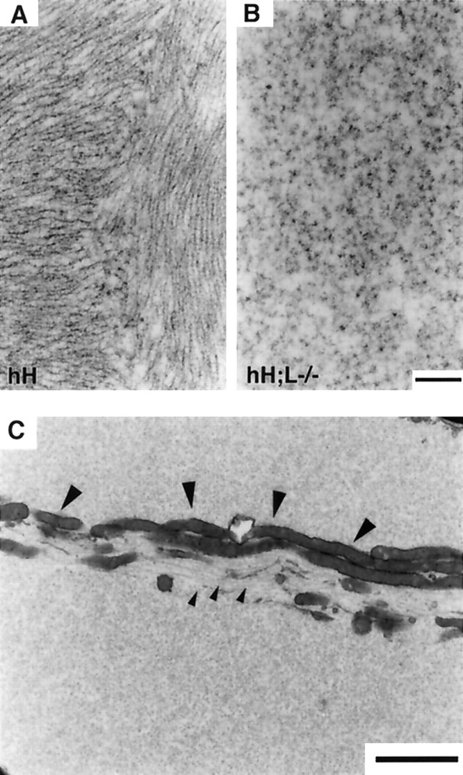
Absence of IF structures in perikaryal inclusions of hH;L−/− mice. A, B, Electron micrographs show IF structures in inclusions of hNF-H transgenic mice (A) and their absence in the inclusions of hH;L−/− mice (B). C, Electron micrograph showing the segregation of microtubules (small arrowheads) and mitochondria (large arrowheads) in an inclusion of hH;L−/− mouse. Scale bars: (in B)A, B, 0.3 μm; C, 2 μm.
