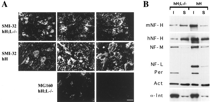
Fig. 4.
The inclusions in hH;L−/− mice are resistant to Triton extraction. Cryostat sections from unperfused 3-month-old hNF-H transgenic and hH;L−/− mice were incubated for 15 min in either PBS or 1% Triton buffer. A double immunofluorescence analysis demonstrates that the NF-H inclusions stained by Smi-32 immunoreactivity (arrowheads) are not extracted by this treatment, whereas the Golgi apparatus membrane-associated protein MG160 is extracted. Scale bar, 50 μm. B, Western blot of soluble (S) and insoluble (I) fractions (2.5 μg) of spinal cord homogenates from 3-month-old hNF-H transgenic (hH) and hH;L−/− mice. Protein detection was performed using the following antibodies: OC95 (1:200), hNF-H; OC59 (1:200), mouse NF-H and NF-M; NN-18 (1:1000), NF-L; NR-4 (1:1000), peripherin (Per); MAB1527 (1:1000), actin (Act); and clone c4 (1:5000) and α-internexin (α-Int) AB1515 (1:2000).

