
Fig. 8.
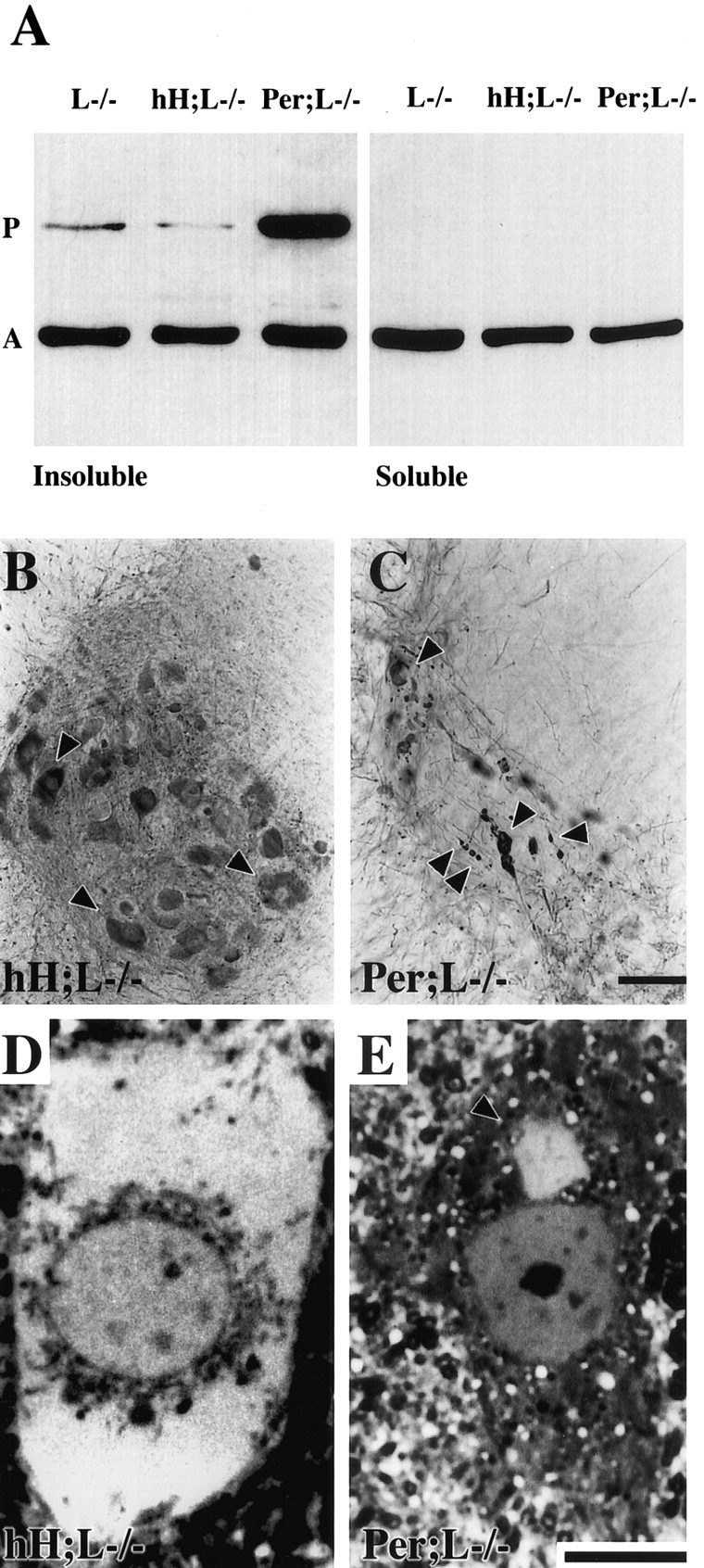
Peripherin detection in protein aggregates of hH;L−/− and Per;L−/− mice. A, Western blot of soluble and insoluble fractions of spinal cord homogenates from NF-L−/− (L−/−), hH;L−/−, and Per;L−/− mice. The insoluble protein fraction (2.5 μ g) and an equal volume of the soluble protein fraction were loaded on gel. Protein detection was performed using the following antibodies: peripherin (P); MAB1527 (1:1000), actin (A); and clone c4 (1:5000). B,C, Immunohistochemical detection of peripherin in the spinal cord with a polyclonal antibody (AB1530, 1:5,000) showing the difference in the size and distribution of peripherin-containing inclusions (arrowheads) between hH;L−/− (B) and Per;L−/− (C) mice. D, E, Thin sections stained with toluidine blue showing a large hyaline inclusion filling the perikaryon of a motor neuron in an hH;L−/− mouse (D) and a typical small hyaline inclusion (arrowhead) in the perikaryon of a motor neuron in a Per;L−/− mouse (E). Scale bars: C, 100 μm;E, 10 μm.