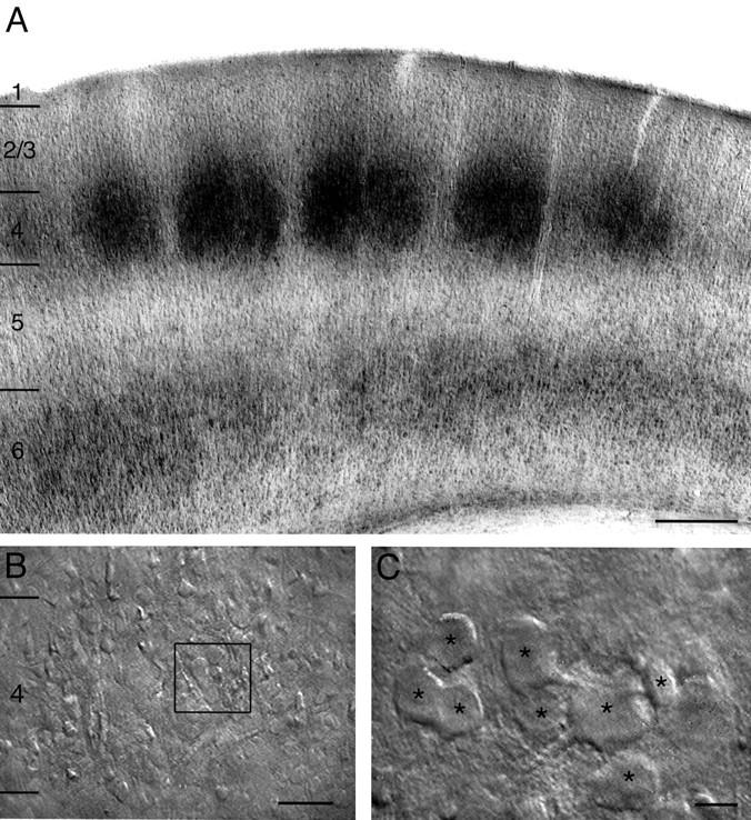
Fig. 1.

A, Low magnification of a semicoronal section through the barrel field (as used for acute slices) stained for cytochrome oxidase showing the regular distribution of barrels in layer 4 of the somatosensory cortex. Scale bar, 500 μm.B, Low magnification IR-DIC contrast image of layer 4 of the barrel cortex. The box outlined inblack indicates a cluster of spiny layer 4 neurons that is shown enlarged in C. Scale bar, 200 μm.C, Higher magnification of a single cluster of spiny layer 4 neurons. All neurons in the plane of focus are marked byblack asterisks. Scale bar, 10 μm.
