
Fig. 3.
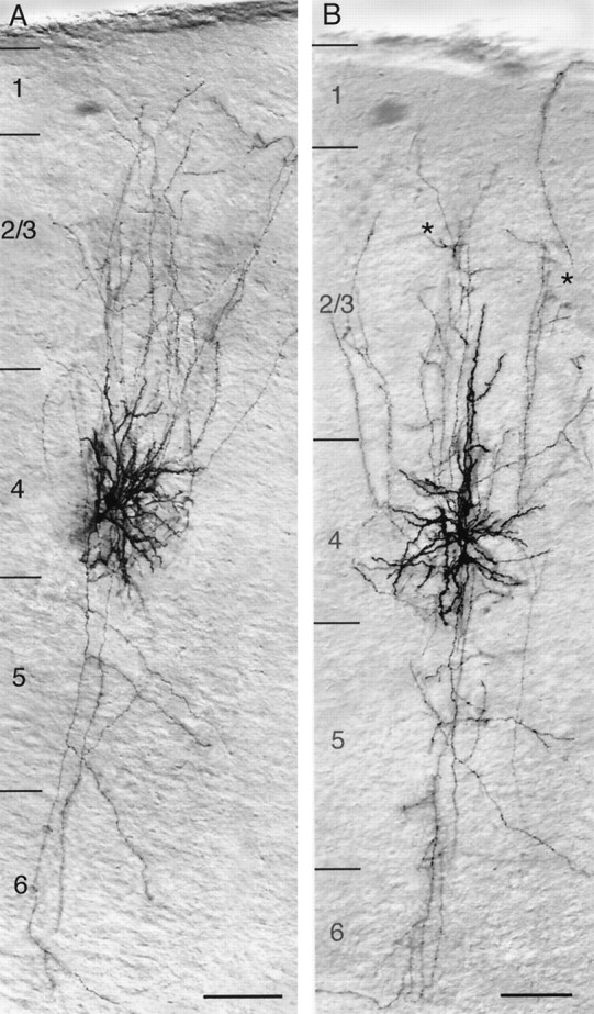
Low-magnification light microscope image of a synaptically coupled pair of spiny stellate cells (A) and star pyramidal neurons (B) filled with biocytin showing the location, dendritic configuration, and columnar axonal projection of both neurons. Note that the characteristic asymmetric dendritic configuration of spiny stellate cells is confined to layer 4, whereas the axons of the presynaptic and postsynaptic neuron project throughout the cortex from layer 1 to the white matter with extensive arborization in layers 2/3 and 4. In contrast, dendrites of star pyramidal neurons have no asymmetric distribution. They are largely confined to layer 4, with the exception of the apical dendrites that terminate in middle to upper layer 2/3. The axons have a projection similar to that of spiny stellate cell axons but tend to show clustering (black asterisks) in layer 2/3. Scale bar, 100 μm.