Fig. 5.

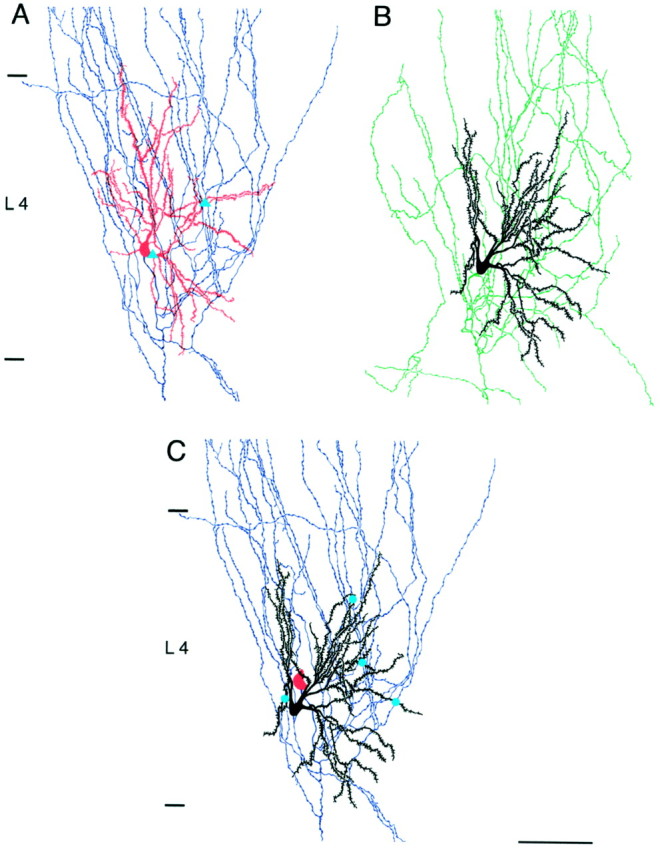
Camera lucida reconstruction of the same pair of spiny stellate cells as shown in Figures 3A and 4.A, The projecting neuron (cell 1) with its dendritic arbor in red and the axon in blue.B, The target neuron (cell 2) with its dendritic arbor in black and the axon in green. Putative autaptic contacts between axon and dendrites of the presynaptic cell are marked with blue triangles. C, Dendritic arbor of cell 2 (target neuron) and axonal projections of cell 1 (projection neuron). Blue dots indicate putative synaptic contacts between the presynaptic axon and the dendrites of the postsynaptic spiny stellate cell as identified by high-power light microscopic examination. The soma of the presynaptic cell is shown inred. Autaptic contacts are not marked inC. Scale bar, 100 μm.
