
Fig. 3.
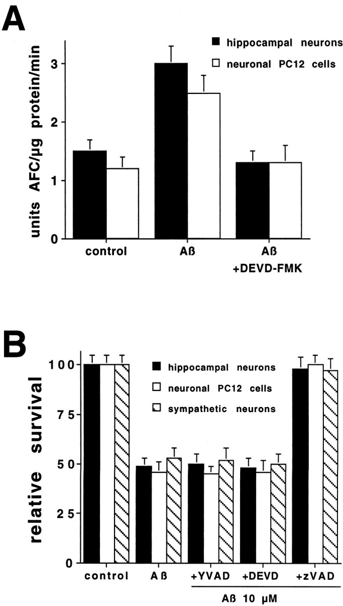
A, Aβ1–42 induces caspase activity in hippocampal neurons and PC12 cells. Hippocampal neurons and neuronal PC12 cells were treated with Aβ1–42(10 μm) with or without DEVD-FMK (10 μm) for 6 hr. Cells were lysed, and 25 μg of protein of each treatment was incubated with the fluorogenic substrate DEVD-AFC (15 μm). The release of AFC was quantified in an LS50B fluorometer. This is a representative experiment; comparable results were obtained in three additional independent experiments.B, Differential protection by caspase inhibitors from Aβ1–42-induced death. Cultures of hippocampal neurons, PC12 cells, and sympathetic neurons were exposed to Aβ1–42 (10 μm) in the presence or absence of the indicated inhibitors (n = 3): YVAD-FMK at 100 μm, DEVD-FMK at 10 μm, and zVAD-FMK at 50 μm. Cells were counted after 1 d as described in Figure 1. Survival is reported relative to untreated cultures and is given as mean ± SEM.