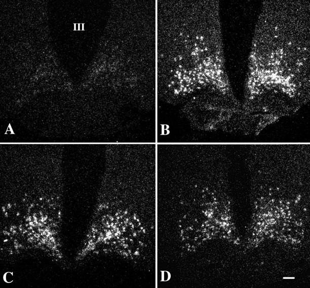
Fig. 4.

Dark-field illumination micrographs of AGRP mRNA in the arcuate nucleus of fed (A) and fasted (B) animals and fasted animals receiving an intracerebroventricular infusion of α-MSH at a dose of 150 ng (C) or 300 ng (D). Note the marked increase in AGRP mRNA in the fasted animals. No significant alteration in AGRP mRNA levels is apparent after α-MSH administration in any of the groups. III, Third ventricle. Scale bar, 100 μm.
