
Fig. 2.
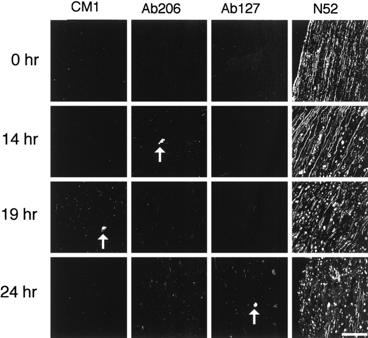
Confocal immunofluorescence micrographs showing that procaspase-3 and its close relatives are not activated during Wallerian degeneration of sciatic nerve. Longitudinal sections of uncut sciatic nerve (0 hr) or sciatic nerve explants cultured for 14, 19, or 24 hr were stained with the N52 anti-neurofilament antibody and with the CM1, Ab206, or Ab127 antibodies. Note the lack of activated caspase-3 in nerves undergoing Wallerian degeneration, even though neurofilaments have been degraded. Arrows point to apoptotic glial cells containing activated caspase-3 or its close relatives. Scale bar, 50 μm.